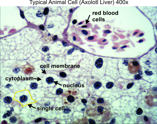
Animal Cell Under Microscope 400x
They are all typical elements of a cell. The cells do not have a cell wall 10.figure 6 shows animal cells from a beef sample stained at 400x
Amazing 27 Things Under The Microscope Diagrams And Descriptions
You can observe this epithelial animal cell under microscope with high power.

Animal cell under microscope 400x. Fresh red blood cells in urine specimen under microscope 400x virtual microscope helps fight against deadly diseases story solved activity 5 osmosis in an animal cell materials fo Light micrograph of a mosss leaf cells at 400x. Phases of the cell cycle.
Title your drawing, spirogyra cell. The granulated area is the cell cytoplasm while the huge round part is the nucleus. A focus on a chain of spirogyra cells under loox.
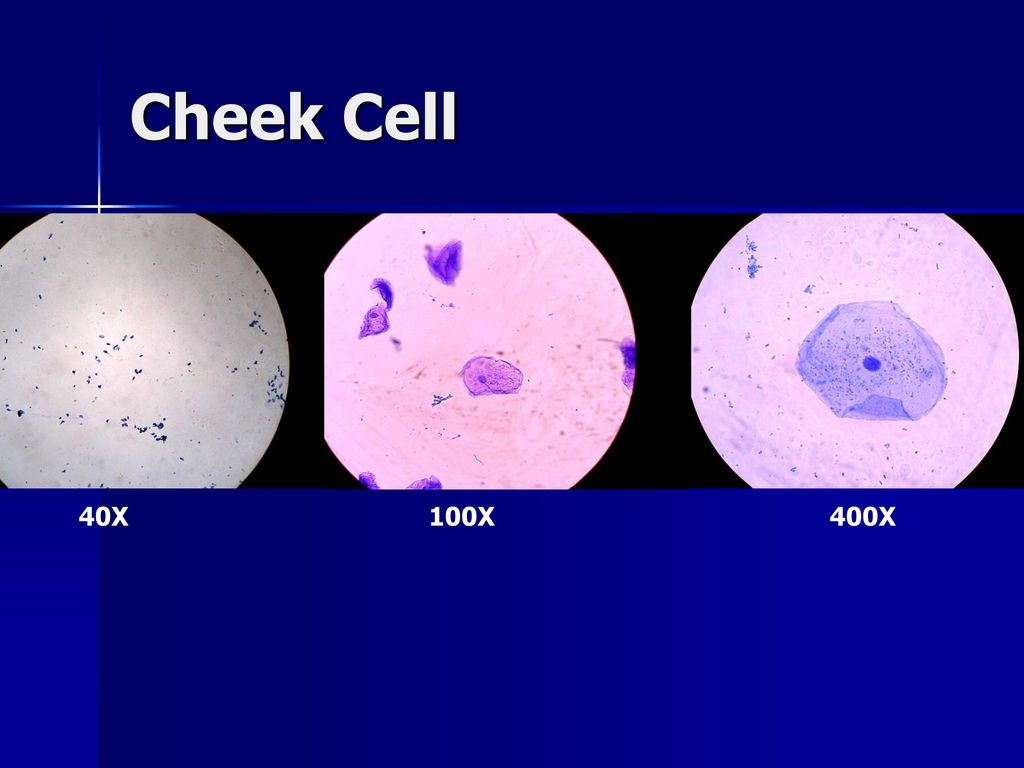
This show consists mainly of clips taken from my microscope and an explanation of. Human cheek cells are made of simple squamous epithelial cells, which are flat cells with a round visible nucleus that cover the inside lining of the cheek.c. Hi, i am yanika (13) and this is my new show 'so microscopic'.
Onion cells under the microscope 40x 100x 400x youtube. Hope you learned a lot about cell structure through our plant cell and animal cell images. Alter observing spirogyra through the microscope, discuss with your partner how you think it got its name.
Microscopic images of plant and animal cells google search. My blood red and white blood cells under microscope 400x clip virtual microscope helps fight against deadly diseases story 400x peripheral area of the lesion with intense red blood cell Click images to large view human skin cells under microscope 400x labeled micropedia.
Click images to large view comparing plant and animal cells microscopy4kids. Draw the cell in the circle on student sheet 7.1: There are two categories of cells, eukaryotic and prokaryotic.
Move your slide so that your field of view is centered on the root tip. Animal cell under light microscope: The plasma membrane should be distinct as a dark border around a light colored cytoplasm.
Focus at 100x and re center so that you are focused on the more 'square' meristem cells. 8.figure 4 shows a typical animal cell structure. Beneath a plant cell’s cell wall is a.
Cheek cell 400x dark field dark field microscopy is a tech flickr. A cell is the smallest functional and structural entity of life that it is easier observing animal cell under light microscope. Since objects viewed under the microscope are very small, the micrometer is used in making such measurements.
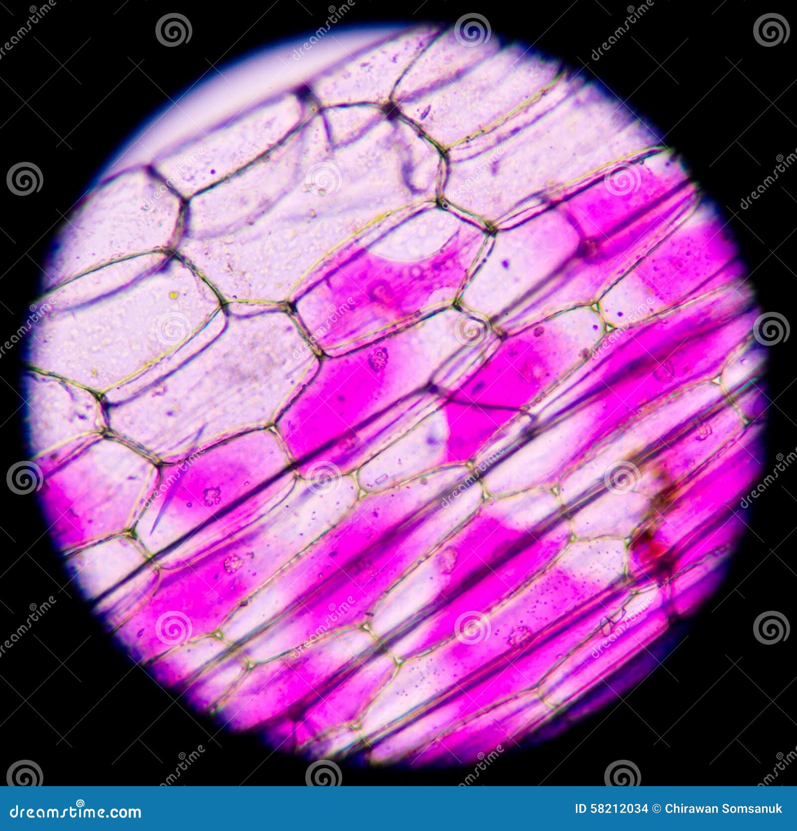
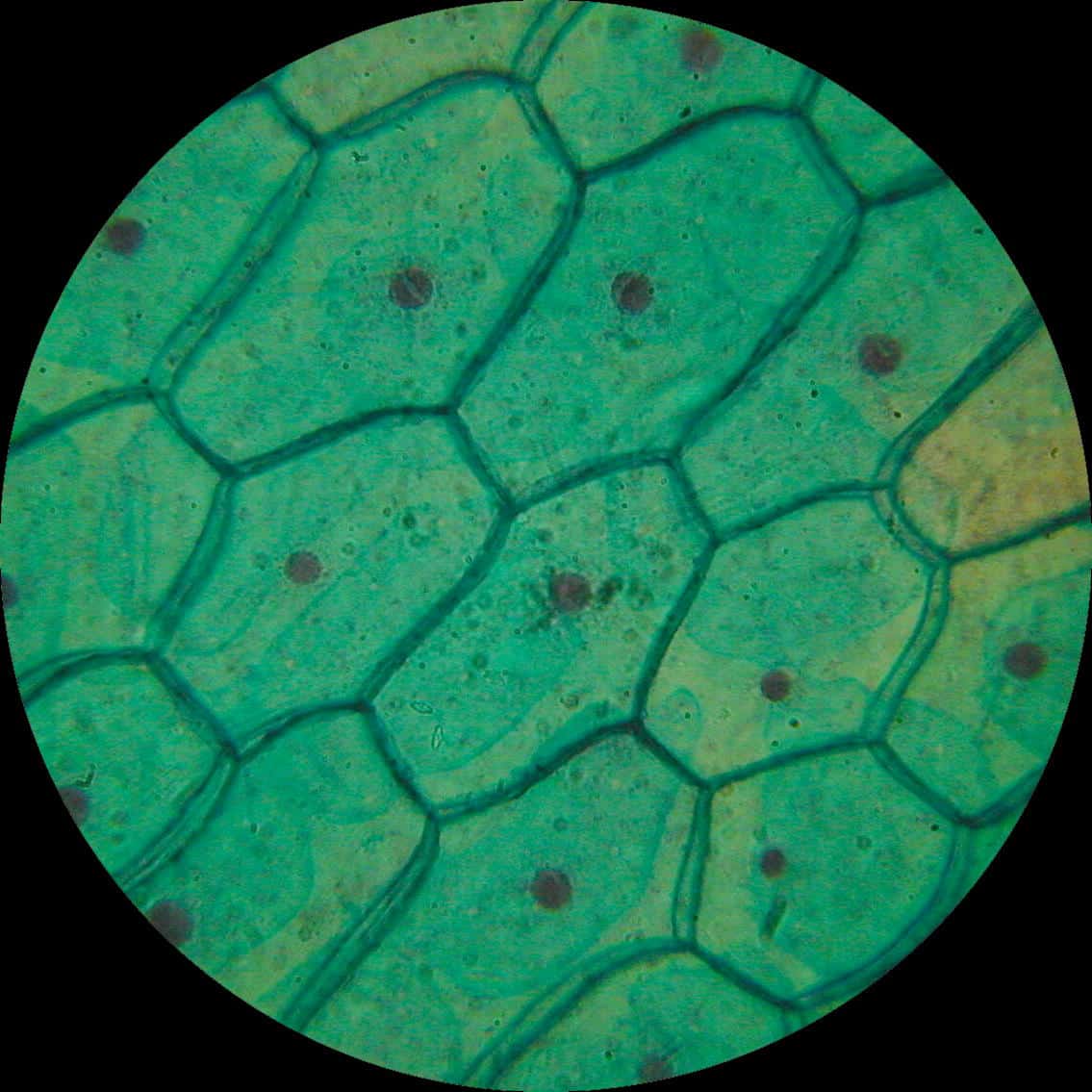
Beneath a plant cell’s cell wall is a cell membrane. Xerophytic leaf c s epidermis plants leaves. C switch to 400x and focus on one cell (see figure 7.1).
At 400x, chloroplasts should be clearly visible in. Cells microscope stock photos cells microscope stock images alamy comparing animal and plant cell microscope lab ppt video online 400x. Cell wall, nucleus and chloroplasts can be seen with a compound light microscope under a total magnification of 400 x.
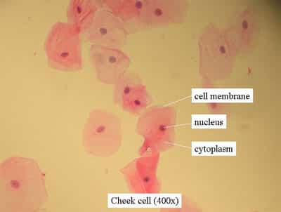
Using a microscope to view the phases of mitosis overview in this exercise you will explore the stages of mitosis using the bionetwork virtual microscope to visualize and identify each stage in both onion root tip and whitefish blastula slides. At 400x, nuclei should be visible in human cheek cells, but no other organelles. In this under the microscope video we are going to see blood (mines) in the microscope in 3 magnifications (40x 100x and 400x) as well as see how to do it.ma.
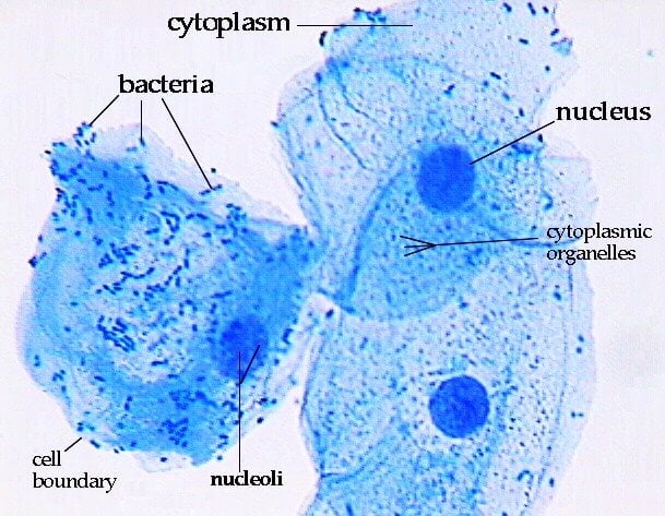
Happy grass cells under a microscope things under a. Click images to large view plant cell lab makeup. 9.figure 5 shows and epithelial cell stained with methylene blue stain at 400x magnification.
There are one or more cells that form organism. At this magnification, flat, irregular shaped cells with thin cell membrane and nucleus can be seen. Generalized cell is used for structure of animal cell and plant cell to present the common parts, appearing in various parts of the bodies of animals and plants.
40x 400x compound monocularbiological microscope45 degree angled headelectric lightedbeginner slides plant cell things under a microscope plant cell picture. Eukaryotic is most complex cells consisting a. Lab activity plant and animal cells under the microscope.
Human cheek cells 400x with images microscopy. Plant and animal cells are alike in that they are both eukaryotic (have a nucleus). Within the cell, there is a shape of round with a circular structure of granulated part on the epithelial cells.
Cells under a microscope by jaimarie nelson. Root anatomy of monocot root biology class anatomy. Plant cell plant cell images things under a microscope.
If you have a microscope 400x and a properly stained slide of the onion root tip or allium root tip you can see the phases in different cells. Comparing plant and animal cells microscopy4kids. Slowly move the slide and search for cells in each phase of mitosis
Set up your microscope, place the onion root slide on the stage and focus on low (40x) power.
Typical Animal Cell Center 100x Stock Photo - Image Of 100x School 152965862
Plant Animal Cells Staining Lab Answers Schoolworkhelper
White Blood Cells In Urine Specimen Under Microscope 400x Stock Photo - Download Image Now - Istock

Plant Cells Under Microscope 400x Stock Photo - Image Of Background Biology 58212034
Microscope Magnification - Ppt Download

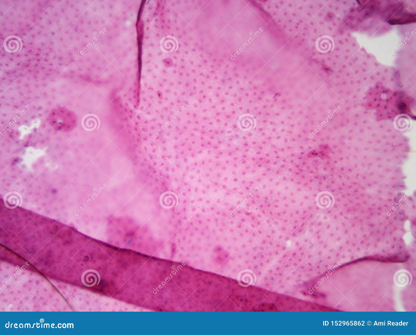
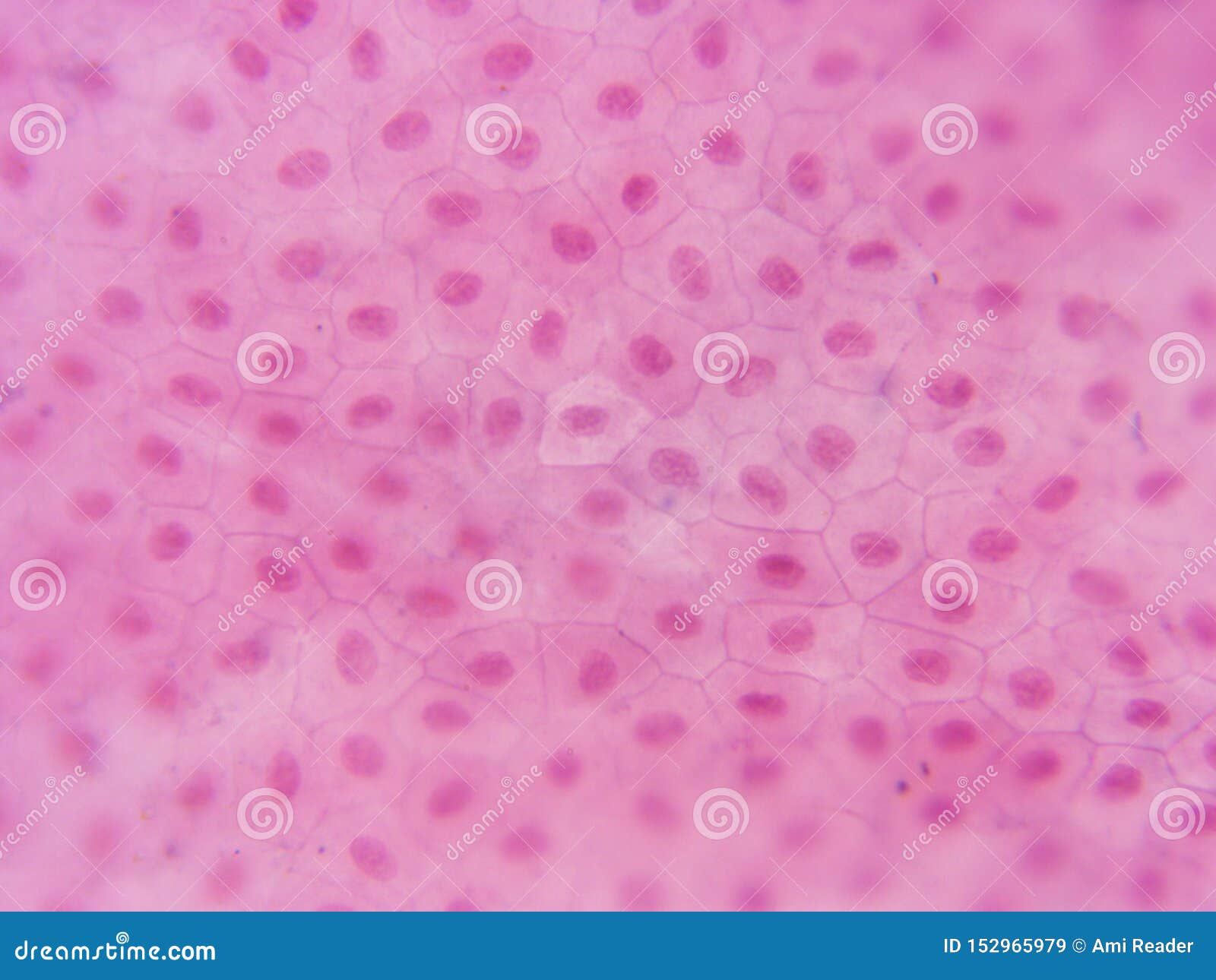
Liver Sections Viewed Under Light Microscope With 400x Magnification Download Scientific Diagram
Pink Plant Cells Under Microscope400x Stock Photo - Download Image Now - Istock


Pin On Microscopic Inspiration For Printmaking Class
Plant Animal Cells Staining Lab Answers Schoolworkhelper
Typical Animal Cell 400x - Dissection Connection

Cheek Cells 400x These Are Cheek Cells Stained With Meth Flickr

Animal Cell Under Microscope Structure And Anatomy

Lab Manual Exercise 1
Typical Animal Cell Center 400x Stock Image - Image Of Visible Compound 152965979
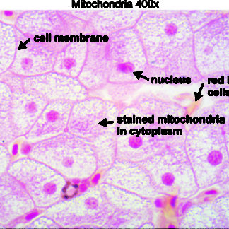
Mitochondria 400x - Dissection Connection
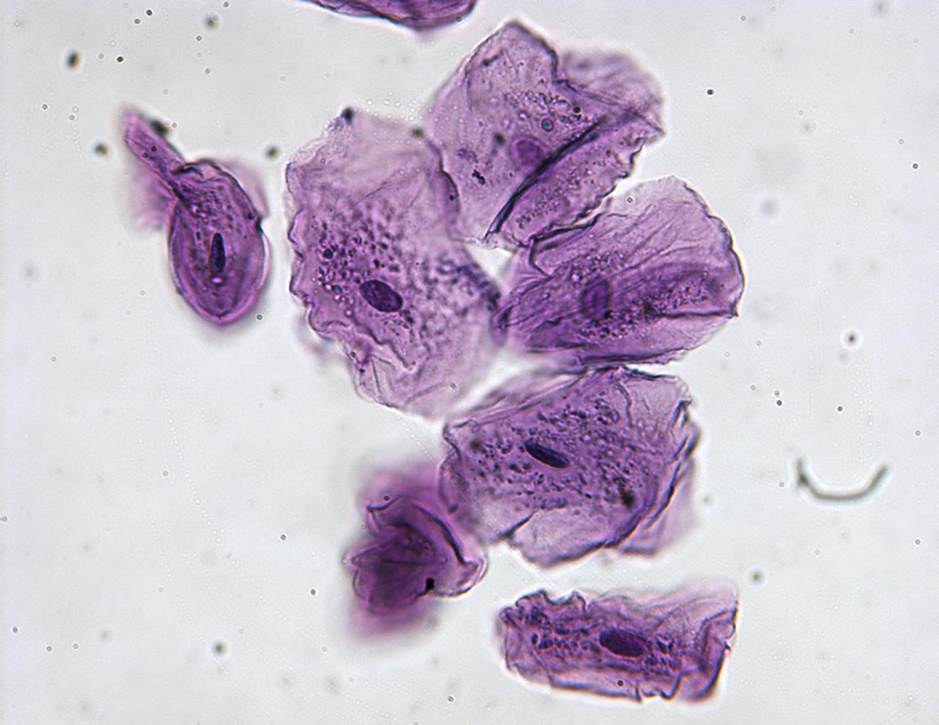
Cheek Cells 400x General Biology Lab Ii Loyola University Chicago

Got My First Setup And Of Course My First Slide Was The Cheek Cell Swab Ive Done Nothing But Make Slides For 2 Days 400x Rmicroscopy
Light Micrographs At 400x Magnification Representative Of The Findings Download Scientific Diagram

Microscopic Animal Cells - Images Kuhn Photo
